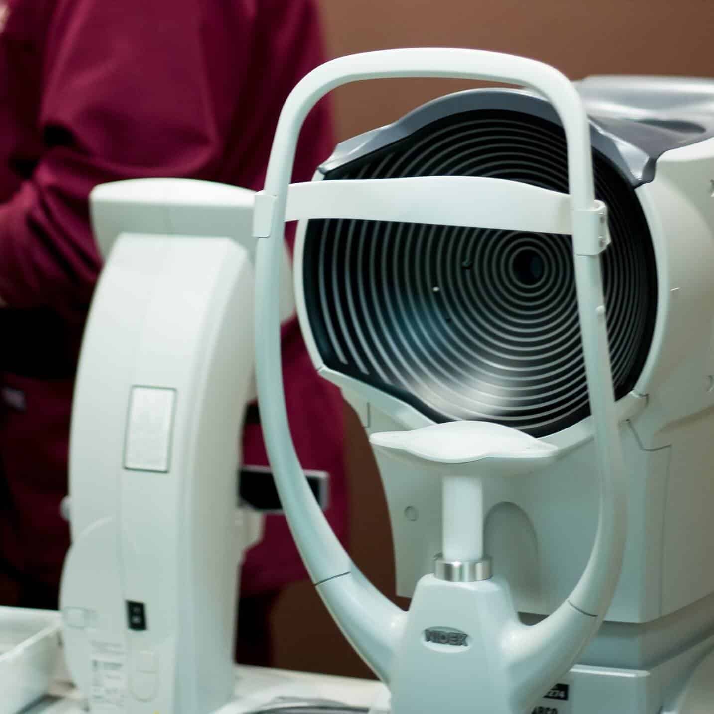
Optical Coherence Tomography
“OCT” is short for “optical coherence tomography”. This is a modern imaging technique like an MRI or sonogram, which shows doctors structures inside the eye that can change due to eye disease. It is a quick and painless test with nothing touching your eye. The images of different areas of the eye will help identify the reason behind impaired vision loss.
In an OCT exam, a light beam scans the eye through the pupil. The beam scans across the back of the eye and the reflected light is translated into a detailed image of the structures within the retina. Careful examination and analysis of the structures seen in these images can help doctors identify early signs of eye diseases like glaucoma and macular degeneration.
OCT has become invaluable in advanced eye care allowing doctors to see very tiny changes in the eye which may otherwise be difficult to detect. This is a tremendous advantage because studies have proven that starting treatment early is the best way to save vision.
Bucks-Mont Eye is proud to have the most advanced OCT equipment on the market today.
Fluorescein Angiography
Fluorescein (Retinal) Angiography is a way of taking pictures of your retina and choroid. The retina is the layer of nerve cells lining the back wall inside of your eye. The choroid is the part of your eye between the sclera and the retina. It contains blood vessels and connective tissue.
Fluorescein Angiography helps in diagnosing certain eye diseases. It also assists the Doctor to track changes over time and target treatment areas. This test is performed in the office and usually takes less than 30 minutes. Bucks-Mont Eye Associates has the latest technology available with our new Heidelberg Spectralis.
During a fluorescein angiogram, you will have drops to dilate your pupils. The Ophthalmologist will inject a colored dye into a vein in your arm. This dye will very quickly travel through your body and as it reaches the blood vessels in your eye, a special camera will take a series of photos. These pictures will be used to determine and guide treatments.
Following this testing, your vision will be blurry for a few hours, so it is wise to have someone drive you. Your skin may also appear a bit yellow for a few hours and your urine may look orange or yellow for up to 24 hours following the angiography. Keep in mind that some people may be allergic to this dye as well. You will want to let your doctor know if you are allergic to things with iodine in them.

